Calan dosages: 240 mg, 120 mg, 80 mg
Calan packs: 30 pills, 60 pills, 90 pills, 120 pills, 180 pills, 270 pills, 360 pills
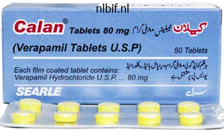
Proven 120 mg calan
In the granular stage blood pressure essentials cheap calan 80mg with visa, the larva continues to degenerate and the cyst develops a hoop enhancement heart attack playing with fire purchase calan 80mg amex. The most definitive neuroim growing older proof of neurocysticercosis is a cystic lesion displaying the scolex arrhythmia in newborns order calan 80 mg visa. Two subjacent small rimenhancing cystic lesions throughout the left parietal juxtacortical white matter hypertension 2 cheap calan 120mg amex. Small rim-enhancing cystic lesion throughout the left parietal juxtacortical white matter. Significant vasogenic edema surrounding the cystic lesion inside the left parietal juxtacorical white matter. In each stage, aside from the vesicular stage, the parasite is within the process of dying. Patients may become symptomatic in the earlier phases, when the parasite elicits an inflammatory response and the lesion becomes surrounded by edema. Prednisone is started either earlier than or with the primary dose of anticysti cidal remedy and continued throughout the course of therapy. Cysticidal drug therapy appears to be most efficacious in sufferers with cysticerci within the colloidal and vesicular stages. There are a quantity of different types of neurosyphi lis, which may be divided into early and late neurosyphilis. Early neurosyphilis includes asymptomatic neurosyphilis, syphilitic meningitis, and meningovascular syphilis. The late forms of neurosyphilis are tabes dorsalis and general paresis (dementia paralytica). Asymptomatic neurosyphilis is outlined by the presence of spinal fluid abnormalities in the absence of neuro logic signs and signs. Syphilitic meningitis is defined by the looks of meningeal indicators and symptoms, including headache, nausea, vomiting, stiff neck, and cranial nerve abnor malities. Spinal fluid evaluation in syphilitic meningitis reveals an increased opening strain, a lymphocytic pleocytosis, a standard or barely decreased glucose con centration, and an elevated protein focus. Meningovascular syphilis is outlined by the appearance of focal neurologic indicators due to an inflammatory arte ritis involving small and mediumsize arteries in asso ciation with signs of meningeal inflammation. General paresis (dementia paralytica) is a persistent pro gressive meningoencephalitis with a peak incidence 10 to 20 years after primary an infection. Tabes dorsalis develops 10 to 20 years after major infection and is characterized at onset by epi sodic lancinating pain within the lower extremities. Due to lumbosacral nerve root dysfunction, decrease extremity areflexia, impotence, and loss of urinary continence could develop. The prognosis of neurosyphilis is made by a combina tion of serologic checks and spinal fluid evaluation. The endemic areas for Lyme illness in the United States are the east coast from New Hampshire to the District of Columbia, parts of Minnesota and Wisconsin, and areas of northern California. Patients with meningitis due to Borrelia burgdorferi complain of headache and fatigue. A unilateral or bilateral facial nerve palsy could also be present or a painful radiculopathy. This lesion is an erythematous lesion that, because it expands, develops central clearing so that it has the appearance of a target lesion. To detect the intrathecal production of antibodies, an antibody index is beneficial. Lyme meningitis, cranial neuritis, and radiculitis is handled with intravenous ceftriaxone for 2 to four weeks. Doxycycline is an inexpensive possibility because it has been used efficiently in Europe for the treat ment of meningitis as a end result of Lyme disease in adults and children 8 years of age or older. Rather, isolated miliary tubercles kind within the brain parenchyma or the menin ges during hematogenous dissemination of bacilli and subsequently enlarge and are often caseating. Subep endymal caseous foci may remain quiescent for months or years but then might discharge bacilli and tuberculous antigens into the subarachnoid area, causing meningi tis. The neurologic complications of tuberculous men ingitis are initiated by the intense inflammatory response to the discharge of tubercle bacilli and tuberculous anti gens into the subarachnoid area. Vasculitis usually entails the major blood vessels at the base of the brain, resulting in cerebral ischemia and infarction.
Buy calan 80 mg on-line
Within one other 3 days arteria iliaca interna order 240 mg calan overnight delivery, by embryonic day 24 blood pressure wrist band generic calan 80 mg visa, the neural tube is closed from the anterior finish (where the mind will form) all through much of the length of the spinal cord heart attack telugu movie online generic 80 mg calan, with the exception of a gap on the rhomboid sinus or posterior neuropore arrhythmia with normal heart rate buy calan 80mg lowest price. At this stage, the neural tube has begun to acquire additional signs of differentiation that reflect the genesis of neurons with distinct features. First, based upon the placement of both the notochord, or the alar area and neural crest, two regions of the neural tube turn out to be specialized to present alerts to the remainder of the neuroepithelial neural stem cells that represent the growing nervous system. The neural tube cells above the notochord at the anterior midline constitute the ground plate, and those on the fusion of the neural folds at the posterior midline turn into the roof plate. Floor plate and roof plate cells secrete signals that influence neighboring cells within the neural tube, such as sonic hedgehog, a peptide hormone that regulates proliferation and differentiation. These geometrically outlined domains of neural stem cells generate functionally distinct lessons of neurons. The neural stem cells of the posterior/alar region will generate sensory projection and interneurons that relay and course of incoming sensory information from peripheral sensory ganglia, and people in the anterior/ basal region will give rise to motor neurons that project to peripheral muscle tissue and autonomic ganglia, as well as interneurons that modulate the output of motor neurons. Signals from the roof plate and flooring plate elicit native expression of transcription factors and other determinants in neighboring neural stem cells. These factors outline the capability of the native stem cells to generate distinct courses of sensory and motor projection or interneurons. These modifications are seen anatomically as the emergences of a sequence of bulges, bends, and grooves that distinguish specific regions of the developing nervous system from the anterior to posterior finish. At the anterior end of the closed neural tube, the neuroepithelium expands right into a hollow globe known as the prosencephalon. The neural stem of the prosencephalon is specified to generate the entire neurons that will comprise the major areas of the forebrain. Subsequently, two bilaterally symmetric structures emerge from the lateral/anterior aspect of the prosencephalon. These are the optic vesicles that may generate the entire neural cells of the retina. Immediately posterior to the prosencephalon, the neural tube bends at some extent referred to as the cephalic flexure. This bending point begins the process by which the mind (and the head) will turn into distinct from the spinal twine and rest of the physique. The stem cells within the neural tube within the area of the cephalic flexure become specified to give rise to the structures of the midbrain (also referred to because the mesencephalon). The region of the neural tube posterior to the midbrain undergoes a dramatic series of morphogenetic modifications that remodel it into the rhombencephalon. The most noticeable event is the institution of a series of repeated bulges and grooves along the anterior/posterior axis that constitute a series of transient domains referred to collectively as rhombomeres. The neural stem cells in each rhombomere purchase distinct patterns of gene expression based mostly upon their location. These distinctions then facilitate native genesis of motor neurons that give rise to the cranial motor Forebrain (prosencephalon) Hypothalamic sulcus Midbrain (mesencephalon) Mesocele Sulcus limitans Hindbrain (rhombencephalon) Rhombocele Alar (roof) plate Sulcus limitans Prosocele Opening of right optic vesicle Basal plate Spinal twine Optic vesicle Forebrain Prosocele Alar (roof) plate Sagittal part Midbrain Mesocele Hindbrain Rhombocele Basal plate Spinal cord Frontal section (anterior to sulcus limitans) Alar (roof) plate Basal plate Derivatives of neural crest nerves, and to sensory neurons that present the targets for peripheral cranial sensory inputs to the brainstem (including the cerebellum/pons, also known as the metencephalon, and the medulla oblongata, also recognized as the myelencephalon). The relationship between rhombomeres and the creating constructions of the pinnacle is type of precise. Similarly, cranial ganglia derived from neural crest that migrates from distinct rhombomeres have a specific relationship with target nuclei generated inside the related rhombomere. The prosencephalon turns into further subdivided into two telencephalic vesicles (collectively called the telencephalon) that can give rise to the bilaterally symmetric constructions of the forebrain: the cerebral cortical hemispheres, the hippocampi, the basal ganglia, basal forebrain nuclei, and the olfactory bulbs. The the rest of the prosencephalon, posterior to the telencephalic vesicles, turns into the diencephalon, which will generate the epithalamus (dorsal structures known as the habenula), thalamus (the relay nuclei that project to the cerebral cortex), and hypothalamus (motor/endocrine control nuclei that regulate visceral and reproductive perform and homeostasis). The mesencephalon, rhombencephalon, and myelencephalon turn into additional differentiated, and the cranial motor nerves (see darker blue within the upper panel of Plate 1-4), sensory ganglia, and associated cranial sensory nerves (lighter pink, Plate 1-4) turn out to be clearly visible along the anterior to posterior extent of the midbrain and hindbrain. In parallel, the motor nerves and sensory ganglia and related sensory nerves of the the rest of the physique turn out to be seen alongside the anterior to posterior extent of the spinal cord. While the neural tube is buying additional regional id that prefigures the ultimate era of the mature neurons and glia in distinct brain areas, the space enclosed by the neural tube turns into further defined as the ventricular system. Within 8 days, the ventricular system has become more elaborate, in parallel with the elaboration of the forebrain, midbrain, and hindbrain. There at the moment are two lateral ventricles enclosed by the telencephalic vesicles, a diocele that can turn out to be the third ventricle, a mesocele that will turn into the cerebral aqueduct, and a metacele and myelocele that can collectively develop into the fourth ventricle. The ventricular space enclosed by the creating spinal twine is now outlined because the central canal.
Syndromes
- Genetic conditions, such as Riley-Day syndrome or Rett syndrome
- Chloroquine
- Avoiding caffeine (coffee, tea, and chocolate)
- Meningitis
- Cartilage repair or resurfacing procedures
- Methods to cause vomiting
- CT scan of the abdomen
- Constipation
Buy 120mg calan with mastercard
Each base subsequently has a complementary base prehypertension at 30 purchase calan 120mg with visa, and the sequence of bases on one strand implies the complementary sequence on the opposite strand blood pressure chart in canada buy 120 mg calan with amex. Polymerase enzymes use the power of this triphosphate to catalyze formation of a phosphodiester bond with the hydroxyl group hooked up to the 3 carbon of the extending strand blood pressure 60100 buy generic calan 240 mg line. Whole genome sequencing will become extra widely used as its cost and evaluation time rapidly lower heart attack 85 year old buy generic calan 240 mg on-line. Translation, folding, modification, transport, and sometimes cleavage to create an lively form of the protein are all regulated steps within the expression of protein-coding genes. Epigenetic adjustments represent a technique by which the surroundings influences gene expression and are necessary for a variety of mobile processes, including improvement, differentiation, and genomic stability. This layer of gene expression control is assumed to regulate the genome to silence aberrant transcripts, buffer fluctuations, and in any other case maintain a certain mobile status quo in response to environmental and cellular cues. Deaminationofcytosineorof its 5-methyl spinoff produces a pyrimidine able to pairing with adenineratherthanguanine. Deamination of cytosine can be accelerated by some mutagenic chemicals such as hydrazine. Ultraviolet gentle causes photochemical dimerization of adjoining thymine residues that will then be altered during restore or replication; in people, that is extra related to somatic mutations in uncovered skin cells than to germline mutations. Movement of such parts or recombination amongst them is a source of spontaneous insertions and deletions, respectively. An amorphic (or null) allele confers a whole loss of operate, a hypomorphic allele confers a partial lack of function, a hypermorphic allele confers a achieve of normal operate, a neomorphic allele confers a achieve of novel perform not encoded by the conventional gene, and an antimorphic or dominant negative allele antagonizes regular operate. The practical impression of allele courses is that distinct medical syndromes may be attributable to completely different alleles of the same gene. For example, totally different allelic mutations in the androgen receptor gene have been tied to partial or complete androgen insensitivity9 (including hypospadias and Reifenstein syndrome), prostate most cancers susceptibility, and spinal and bulbar muscular atrophy. Approximately 2% to 5% of the human genome encodes protein or confers regulatory specificity. Given present estimates of roughly 25,000 human genes2,three and provided that more than 7 billion people inhabit the earth, one might anticipate that every human carries one or more mutations. Several public databases that curate details about human genes and mutations are available online (Table 1-1). In addition to the nuclear chromosomes, the mitochondrial genome contains roughly 37 genes on a round unit that resides in this organelle, in anyplace from a hundred to one hundred,000 copies. Five major teams of histones are essential for correct packing of chromatin, and the heterogeneous nonhistone proteins are required for regular gene expression and higher-order chromosome packaging. The nucleosome "beads" are further condensed into higher-order structures referred to as solenoids, which can be packed into loops of chromatin that are attached to nonhistone matrix proteins. This orderly packing permits each chromosome to be faithfully wound and unwound throughout replication and cell division. Additionally, chromatin group plays an essential role within the management of gene expression. In the human, there are forty six chromosomes in a traditional cell: 22 pairs of autosomes and two intercourse chromosomes, X and Y (see later discussion). Each chromosome accommodates a centromere, a constricted region that varieties the attachments to the mitotic spindle and governs chromosome movements during mitosis. The chromosomal arms radiate on all sides of the centromere and terminate within the telomere or finish of every arm. The means of cell division is identified as mitosis, and the interval between divisions is known as interphase. Mitosis is a steady course of that could be arbitrarily divided into four stages based on the morphology of the chromosomes and the mitotic equipment. The starting of mitosis is characterised by swelling of chromatin, which becomes seen underneath a light microscope by the end of this prophase. When a cell is in metaphase or is coming into metaphase from prophase, clinical strategies to study chromosomes trigger arrest of additional steps in mitosis. Therefore, all sister chromatids (4n) are visible in a regular clinical karyotype.
Calan 80 mg visa
The parietooccipital sulcus is situated primarily on the medial surface of the cerebrum pulse pressure 37 safe calan 240 mg, nevertheless it cuts the superior margin and seems for a short distance on the superolateral floor about 5 cm in entrance of the occipital pole hypertension lowering foods discount calan 120mg on-line. The above options divide the cerebrum into frontal blood pressure chart by age nhs buy calan 120 mg free shipping, parietal blood pressure cuff and stethoscope purchase 80 mg calan amex, occipital, and temporal lobes. The frontal lobe lies in front of the central sulcus and anterosuperior to the lateral sulcus. The parietal lobe lies behind the central sulcus, above the posterior ramus of the lateral sulcus and in entrance of an imaginary line drawn between the parieto-occipital sulcus and the preoccipital notch. Parietal lobe Frontal lobe Occipital lobe Occipital pole Calcarine fissure Lunate sulcus (inconstant) Transverse occipital sulcus Preoccipital notch Inferior (inferolateral) margin of cerebrum Inferior temporal gyrus Temporal lobe Central sulcus of insula Circular sulcus of insula Insula Short gyri Limen Long gyrus the occipital lobe lies behind this same imaginary line. The temporal lobe lies below the stem and posterior ramus of the lateral sulcus, and is bounded behind by the decrease part of the aforementioned imaginary line. The superolateral surface of the frontal lobe is traversed by three primary sulci and thus divided into 4 gyri. The precentral sulcus runs parallel to the central sulcus, separated from it by the precentral gyrus, the good cortical somatomotor space. The superior and inferior frontal sulci curve throughout the remaining a half of the floor, dividing it into superior, center, and inferior frontal gyri. The postcentral sulcus lies parallel to the central sulcus, separated from it by the postcentral gyrus, the great somatic sensory cortical space. The outer surface of the occipital lobe is less intensive than that of the other lobes and has a short transverse occipital sulcus and a lunate sulcus; the latter demarcates the visuosensory and visuopsychic areas of the cortex. The temporal lobe is divided by superior and inferior temporal sulci into superior, middle, and inferior temporal gyri. The sulci run backward and barely upward, in the same basic course because the posterior ramus of the lateral sulcus, which lies above them. The superior sulcus ends in the lower a part of the inferior parietal lobule, and the superjacent cortex is called the angular gyrus. The insula is a sunken lobe of cortex, overlaid by opercula and buried by the exuberant development of adjoining cortical areas. It is ovoid in shape and is surrounded by a groove, the round sulcus of the insula. The apex is inferior, near the anterior (rostral) perforated substance, and is termed the limen of the insula. The insular floor is split into larger and smaller posterior components by the central sulcus of the insula, which is roughly parallel to the central sulcus of the cerebrum. The corpus callosum is the most important of the cerebral commissures, and varieties most of the roof of the lateral ventricle. In a median sagittal part, it appears as a flattened bridge of white fibers, and its central part, or trunk, is convex upward. The anterior finish is recurved to type the genu, which tapers rapidly into the podium. Below the splenium and trunk of the corpus callosum are the symmetric arching bundles (crura of the fornix) that meet to form the body of the fornix and separate again to become the columns of the fornix, curving downward to the mammillary our bodies. The cingulate sulcus is easily identified on the medial surface, mendacity parallel to the corpus callosum. It begins beneath the genu of the corpus callosum and ends above the posterior part of the trunk by turning upward to minimize the superior margin of the hemisphere. Opposite the middle of the trunk is another vertical department sulcus, and the world of cortex between these ascending sulci is the paracentral lobule, which accommodates parts of the motor and sensory cortical areas. The cingulate sulcus separates the medial frontal and cingulate gyri, and below the genu and rostrum of the corpus callosum are small parolfactory sulci separating the subcallosal (parolfactory) areas and paraterminal gyrus. The upper parietooccipital sulcus inclines backward and upward to cut the superior border. The decrease calcarine sulcus extends forward from the occipital pole to end beneath the splenium of the corpus callosum, and the isthmus of cortex between them connects the cingulate and parahippocampal gyri. The wedge-shaped region between the parietooccipital and calcarine sulci is the cuneus, while the world between the parietooccipital sulcus and the paracentral lobule is the precuneus. The major visuosensory area is located within the partitions of the calcarine sulcus and in the adjacent cortex.
Discount calan 80mg
The fastigial nucleus connections with the contralateral inferior olivary nucleus are within the caudal a part of the medial accent olive arteria en ingles buy cheap calan 120mg. Fastigial nucleus efferents affect a quantity of func tional domains: axial and limb girdle musculature (medial motor system) by way of the vestibular and reticular nuclei; oculomotor techniques blood pressure 5020 buy 120 mg calan free shipping, including vertical and horizontal gaze facilities within the midbrain and pons; autonomic facilities via connections with brainstem and hypothala mus; and emotional modulation through links with limbicrelated circuits pulse pressure pda generic 120 mg calan free shipping. They provide cer ebellar efferents in the superior cerebellar peduncle from the predominantly motorrelated spinocerebel lum that receives proprioceptive and exteroceptive inputs from the spinal cord and brainstem blood pressure when to go to er 80 mg calan with visa, and senso rimotor information from the cerebral cortex. This purple nucleus sector supplies the origin for rubrospinal fibers that act on the spinal motor apparatus, particularly arm and hand flexor muscle tissue. Multiple different brainstem connections of the inter positus nuclei include (1) the lateral reticular nucleus and medullary reticular formation giving rise to reticulospinal tracts, (2) the vestibular nuclei as source for vestibulospinal tracts, (3) the superior colliculus giving rise to the tectospinal tract, (4) the oculomotor nuclei (prepositus hypoglossi, Darkschewitsch, and posterior commissures), (5) the sensory (lateral/external cuneate nucleus), and (6) the nociceptive systems (periaqueduc tal grey, medullary raphe). These rostrally directed fibers from the interpositus nuclei continue to the hypothalamus and zona incerta earlier than reaching the thalamus. The dorsal part (paleodentate, due to its relation ship to the paleo, or spinocerebellum) is linked with motor areas of the cerebral cortex. The ventral part (neodentate, interconnected with the extra just lately evolved neocerebellum) is linked with cerebral associa tion areas. Dorsal dentate nucleus fibers terminate in motorrelated thalamic nuclei, together with the ventroposterolateral and ventral lateral nuclei, that then project to the primary motor and pre motor cerebral cortex. Middle and caudal thirds of the dentate nucleus are linked through the ventral anterior nucleus of thalamus with the premotor cortex and with the frontal eye fields engaged in saccadic eye movements. Ventral and lateral parts of the dentate nucleus project via the dorsal sector of the ventral lateral nucleus and the medial dorsal nucleus to dorso lateral prefrontal, posterior parietal, and other cerebral association areas. Dentate nucleus projections to tha lamic intralaminar nuclei provide widespread influence on cerebral cortical areas. These intralaminar nuclei additionally project to the striatum, offering an indirect hyperlink between cerebellum and basal ganglia. The dentate nucleus also tasks to the smallcelled (parvicellular) a half of the pink nucleus that feeds back through the central tegmental tract to the inferior olive, which, in turn, is linked with the cerebellum. The rostral and dorsomedial elements of the dentate nucleus (the paleodentate) project to the dorsal lamina and bend of the principal olive. The ventral and caudal elements of the dentate nucleus (the neodentate) project to the ventral lamina of the principal olive. Lesions of these different pathways produce a broad selection of impairments, motor and in any other case. The vestibular system is also associated to the paleocerebellum (spinocerebellum) through its connections with anterior lobe vermis and the fastigial nucleus. Together with the perihy poglossal nucleus, they also project to the fastigial nucleus as collaterals of vestibulocortical fibers. Axons of each pathways terminate as diffusely projecting mossy fibers in granule cell glomeruli within the cere bellar cortex. These olivary nuclei obtain inhibitory enter from the parasolitary nucleus that, in flip, receives projections from the labyrinth. The olivocerebellar terminations are organized in dis crete parasagittal zones, relaying information from the vertical and anterior semicircular canals. The other branch terminates with varying levels of inten sity in several subregions of all 4 vestibular nuclei: medial, lateral, superior, and inferior. Strong topographically organized projections to the vestibular nuclei are additionally derived from the fastigial nucleus. The rostral fastigial nucleus, linked with the spinal recipient anterior vermis, tasks to the medial, superior, and perihypoglossal nuclei. The caudoventral region of the fastigial nucleus, devoted to oculomotor control, proj ects to the inferior vestibular nucleus and to the a half of the lateral vestibular nucleus that receives zone B corti cal inputs. The projections of the rostral portion of the fastigial nucleus are ipsilateral, whereas fibers from the caudal fastigial nucleus cross in the hook bundle of Russell to excite contralateral vestibular neurons. Thus the vestibular system is crucial for the management of eye actions for orientation in intrapersonal and further private space and for control of the axial musculature, important for steadiness.
Discount 240mg calan
The lateral (sylvian) sulcus has a brief stem between the orbital floor of the frontal lobe and the temporal pole; in life blood pressure 400 generic calan 120 mg with amex, the lesser wing of the sphenoid bone initiatives into it arrhythmia recognition test calan 120 mg with visa. At its outer end arterial thrombosis buy cheap calan 120 mg on-line, the stem divides into anterior blood pressure pills kidney failure 240mg calan visa, ascending, and posterior branches. These rami separate triangular areas of cortex known as opercula, which cover a buried lobe of cortex, the insula. The central (rolandic) sulcus proceeds obliquely downward and ahead from a degree on the superior border nearly midway between the frontal and occipital poles. It is sinuous and ends above the middle of the posterior ramus of the lateral sulcus. Its higher end normally runs onto the medial floor of the cerebrum and terminates in the paracentral lobule. The orbital floor rests on the roofs of the orbit and nose and is marked by an H-shaped orbital sulcus, in addition to by a straight groove on the medial side, the olfactory sulcus, which lodges the olfactory bulb and tract. The orbital sulcus demarcates the orbital gyri; the small convolution medial to the olfactory sulcus is the straight gyrus. The tentorial surface lies partly on the floor of the center cranial fossa and partly on the tentorium cerebelli. Both run nearly instantly ahead from the occipital pole to the temporal pole; like different sulci, they may be subdivided, and the Lateral occipitoPulvinar temporal gyrus Red nucleus OccipitoSuperior (cranial) colliculus temporal Medial occipitotemporal gyrus sulcus Cerebral aqueduct (of Sylvius) Collateral sulcus Splenium of corpus callosum Parahippocampal gyrus Lingual gyrus Apex of cuneus Uncus Occipital pole Calcarine sulcus Cingulate gyrus Cerebral longitudinal fissure anterior end of the collateral sulcus known as the rhinal sulcus. The dentate gyrus, a slender fringe of cortex with transverse markings, occupies the groove between the parahippocampal gyrus and the fimbria of the hippocampus. The anterior end of the parahippocampal gyrus becomes recurved to kind the uncus, which is partly occupied by the cortical olfactory area. The medial occipitotemporal gyrus is fusiform in shape, and lies between the collateral and occipitotemporal sulci. The lateral occipitotemporal gyrus lies lateral to the occipitotemporal sulcus and is steady with the inferior temporal gyrus around the inferior margin of the hemisphere. The cerebral cortex has definite areas related to specific neurologic functions, either for primary sensory reception or for complicated built-in exercise. This is as a result of of the rapid activity along a giant number of precisely organized, reciprocally performing affiliation pathways. The pathways could additionally be very quick, linking neighboring areas and working solely within the gray matter, or they might be longer (arcuate) bundles, passing via the white matter to join gyrus to gyrus or lobe to lobe within a cerebral hemisphere� intrahemispheric connection. Other commissural bundles conduct interhemispheric exercise: probably the most prominent are the corpus callosum, a large band of fibers, which lies instantly beneath the cingulum; the anterior commissure, which connects both temporal lobes; and the hippocampal commissure (commissure of the fornix), which connects the best and left hippocampus. The reciprocal exercise of the connections in the cerebral cortex ensures the coordination of sensory enter and motor exercise, as nicely as the regulation of higher perform. For instance, for the appreciation and integration of visual information, the first visual sensory area of the occipital cortex is linked to the visible affiliation areas. These visible facilities are related by intrahemispheric fibers to the ipsilateral parietal cortex, as properly as to different areas, such because the temporal lobe, for further built-in activity. The right and left parietal and posterior temporal areas, in flip, are connected by the corpus callosum. The prefrontal cortex, (which includes the three frontal gyri, the orbital gyri, many of the medial frontal gyrus, and approximately half of the cingulate gyrus) is concerned with larger psychological functions, and is involved with many behavioral features of man. This space receives quite a few connections from the temporal and parietal lobes by way of pathways in the cingulum, a bundle of long association fibers mendacity within the cingulate gyrus. Bilateral lesions of the prefrontal space produce a lack of concentration, a decreased intellectual capability, and reminiscence and judgment deficits. The somatosensory cortex, which occupies contiguous parts of the frontal and parietal lobes, and the premotor cortex of the frontal lobe are involved with the initiation, activation and efficiency of motor exercise, and the reception of main sensation of the physique. Lesions of the somatosensory cortex end in contralateral paralysis and lack of somatosensory reception or perception. Lesions within the parietal lobe result in sensory ataxia, a loss of basic awareness, faulty recognition of sensory impulses, and an absence of interpretation of spatial relationships. Lesions of the striate cortex (the main visible area) on one facet lead to a contralateral hemianopsia, while lesions of the secondary regions of the visual cortex trigger a scarcity of ability to interpret visible impulses. The posterior part of the temporal lobe is concerned with the reception and interpretation of auditory info, and with some elements of pattern recognition and better visible coordination; the interconnections of the auditory and visible segments of the occipital, temporal, and parietal lobes make this a highly integrated operate.
Black Dogwood (Alder Buckthorn). Calan.
- What other names is Alder Buckthorn known by?
- What is Alder Buckthorn?
- Treating cancer.
- Dosing considerations for Alder Buckthorn.
- Treating constipation.
- How does Alder Buckthorn work?
- Are there safety concerns?
Source: http://www.rxlist.com/script/main/art.asp?articlekey=96823

Generic calan 240 mg without prescription
The location and pattern of hemorrhage may be helpful in figuring out the precise web site of the ruptured aneurysm heart attack 720p generic calan 80mg visa, and these findings could also be used to predict the event of vasospasm blood pressure medication and weight loss purchase calan 80 mg fast delivery. Digital subtraction angiography often is carried out to define the aneurysm and the adjoining vasculature heart attack feat sen city buy calan 80mg cheap, significantly to display for the presence of vasospasm heart attack headache generic calan 120 mg on-line. Additional diagnostic research embody an electrocardiogram, coagulation research, full blood depend, renal perform studies, and electrolytes. Aneurysm has been decompressed with surgical clips at its base with preservation of the father or mother artery (arrowheads). Early issues embrace cardiac arrhythmias, pulmonary edema, electrolyte disturbances, acute hydrocephalus, and seizures. The risk of recurrent aneurysmal bleeding could additionally be as high as 4% inside the first 24 hours and 20% in the first 10 days. Patients should be hospitalized in a stroke unit or intensive care unit, with frequent monitoring of vital indicators and neurologic standing. Those individuals having a decline in consciousness or a compromised airway are intubated. Measures to stop or deal with cardiac arrhythmias, hypertension, electrolyte or metabolic disturbances, or other medical problems are instituted. Increased intracranial pressure is managed with placement of a ventricular drain or medications corresponding to hypertonic saline or mannitol. Symptoms such as pain, nausea, vomiting, or agitation ought to be medically handled. Nimodipine is accredited for prevention of cerebral ischemia secondary to vasospasm. Patients who develop ischemic symptoms are treated with hypervolemic hemodilution and induced hypertension, intra-arterial administration of vasodilators, or angioplasty, relying on the severity of the vasospasm. Moyamoya is a progressive occlusive arteriopathy of the distal inner carotid arteries. The idiopathic form, also called "primary moyamoya" or "moyamoya illness," occurs extra commonly in youngsters of Japanese or Korean descent, although it has been observed in all ethnicities. Secondary moyamoya, or "moyamoya syndrome," can develop after mind radiation for the treatment of childhood cancers, mostly retinoblastoma, or can happen in genetic situations, corresponding to sickle cell disease, Down syndrome, neurofibromatosis type 1, and a uncommon form of primordial dwarfism. The name, Japanese for "haze" or "puff of smoke," comes from small collateral blood vessels that form near the positioning of occlusion and provides a hazy look on standard angiography. Moyamoya sometimes manifests with ischemic strokes or transient ischemic attacks in early to mid childhood. However, if a baby develops sufficient collateral blood move to preclude ischemic events, he or she might not present until young adulthood with a hemorrhagic stroke, sometimes due to rupture of the abnormal moyamoya collaterals. Surgical treatment of moyamoya contains a selection of revascularization procedures intended to bypass the inner carotid circulation and enhance cerebral perfusion. The cerebrovascular anomalies differ widely from clinically insignificant "regular variants," such as a duplicated vessel or persistent fetal vessel, to extreme hypoplasia of the internal carotid artery that can lead to ischemic stroke. Findings on examination will include a pulsatile cranial bruit, macrocephaly, and outstanding scalp veins. To avoid dependent edema, affected upper limb supported on pillow with shoulder abducted, hand slightly larger than elbow, and elbow slightly greater than shoulder. Small towel roll or orthosis used to preserve hand in functional position and minimize contractures of finger or wrist. Towel roll alongside trochanteric region and thigh (extending slightly under body to secure it) prevents external rotation of paretic limb. Pressure stockings prevent deep vein thrombosis and thrombophlebitis, which can lead to pulmonary embolism. Such a program is multidimensional and infrequently instituted inside the first 24 to 28 hours. Positioning after stroke is carried out with goals of preventing joint contractures, edema of the paretic extremity, stress ulcers over bony prominences, and aspiration.
Purchase calan 240mg with mastercard
This isozyme is expressed primarily within the pores and skin and contributes to sebaceous gland activity and the zits related to puberty blood pressure medication sleepy discount calan 120 mg. Peripheral Testosterone Actions Fates and Actions of Androgens Intratesticular Androgen the testosterone produced by Leydig cells has a quantity of fates and multiple actions arteria festival 2013 buy 240 mg calan fast delivery. Testosterone levels inside the seminiferous tubules which may be higher than 100 occasions more concentrated than circulating testosterone ranges are completely required for regular spermatogenesis arrhythmia generator purchase 120 mg calan free shipping. It induces growth of the male tract from the mesonephric duct within the absence of 5-reductase blood pressure 7050 buy cheap calan 80mg on line. Hypothalamic-Pituitary-Testicular Axis the testis is regulated by an endocrine axis. Transport and Metabolism of Androgens As testosterone enters the peripheral circulation, it binds to and rapidly reaches equilibrium with serum proteins. Approximately 50% of excreted androgens are found as urinary 17-ketosteroids, with most of the the rest being conjugated androgens or diol or triol derivatives. Only about 30% of the 17-ketosteroids in urine are from the testis; the remainder are produced from adrenal androgens. Androgens are conjugated with glucuronate or sulfate in the liver, and these conjugated steroids are excreted in urine. In addition to stimulating synthesis of proteins concerned in the "nurse cell" facet of Sertoli cell operate. Male Reproductive Tract Once spermatozoa emerge from the efferent ductules, they go away the gonad and enter the male reproductive tract. The segments of the tract are the: epididymis (head, body, and tail), vas deferens, ejaculatory duct, prostatic urethra, membranous urethra, and penile urethra. In addition to conveying sperm, the primary capabilities of the male reproductive tract are: 1. Sperm spend a couple of month in the epididymis, the place they undergo further maturation. The epithelium of the epididymis is secretory and provides numerous components to the seminal fluid. Spermatozoa that enter the top of the epididymis are weakly motile but are strongly unidirectionally motile by the point they exit the tail. Spermatozoa also bear the method of decapacitation, which involves changes in the cell membrane to prevent spermatozoa from undergoing the acrosome response earlier than contact with an egg (see later). Loweredintratesticular testosterone levels lead to reduced sperm manufacturing and might trigger infertility. Sperm are stored in the tail of the epididymis and vas deferens for several months with out loss of viability. The main perform of the vas deferens, apart from providing a storage web site, is to propel sperm during sexual activity into the male urethra. Normally in response to repeated tactile stimulation of the penis during coitus, the muscularis of the vas deferens receives bursts of sympathetic stimulation that trigger peristaltic contractions. Emission instantly precedes ejaculation, which is the propulsion of semen out of the male urethra. During emission, contraction of the vas deferens coincides with contraction of the muscular coats of the two accent sex glands, the seminal vesicles (right and left) and the prostate gland (which surrounds the prostatic urethra). The seminal vesicles also secrete semenogelins, which induce coagulation of semen instantly after ejaculation. The alkaline secretions of the prostate, which make up about 30% of the volume, are excessive in citrate, zinc, spermine, and acid phosphatase. This secretion is excessive in mucus, which lubricates, cleanses, and buffers the urethra. Men with sperm counts under 20 million/mL, lower than 50% motile sperm, or lower than 60% usually conformed sperm are normally infertile. Emission and ejaculation occur during coitus in response to a reflex arc that includes sensory stimulation from the penis (via the pudendal nerve) adopted by sympathetic motor stimulation to the sleek muscle of the male tract and somatic motor stimulation to the musculature related to the bottom of the penis. However, for sexual intercourse to happen in the first place, the person has to achieve and preserve an erection of the penis. The penis has advanced as an intromittent organ designed to separate the walls of the vagina, cross through the potential house of the vaginal lumen, and deposit semen on the distal finish of the vaginal lumen near the cervix.
Order 80 mg calan mastercard
As a result arteria carotis communis purchase calan 120 mg online, sequestration of calcium throughout diastole is enhanced and the comfort time is shortened prehypertension 125 order calan 240mg fast delivery. Increased ryanodine Ca++ channels in the sarcoplasmic reticulum promote release of Ca++ from the sarcoplasmic reticulum during systole blood pressure chart male buy calan 80mg on line. Effects on Basal Metabolic Rate and Thermogenesis Increased O2 use finally is determined by an increased supply of substrates for oxidation 5 fu arrhythmia order calan 120 mg on-line. T3 augments glucose absorption from the gastrointestinal tract and will increase glucose turnover (glucose uptake, oxidation, and synthesis). In adipose tissue, thyroid hormone induces enzymes for the synthesis of fatty acids, together with acetyl-CoA carboxylase and fatty acid synthase, and enhances lipolysis by increasing the variety of -adrenergic receptors (see Effects on the Autonomic Nervous System). Thus lipid turnover (free fatty acid release from adipose tissue and oxidation) is augmented. Protein turnover (release of muscle amino acids, protein degradation, and to a lesser extent protein synthesis and urea formation) is also increased. T3 potentiates the respective stimulatory effects of epinephrine, norepinephrine, glucagon, cortisol, and development hormone on gluconeogenesis, lipolysis, ketogenesis, and proteolysis of the labile protein pool. The overall metabolic effect of thyroid hormone has been aptly described as accelerating the physiological response to hunger. In addition, thyroid hormone stimulates synthesis of bile acids from ldl cholesterol and promotes biliary secretion. The internet impact is a decrease in the physique pool and plasma ranges of complete and low-density lipoprotein cholesterol. Metabolic clearance of adrenal and gonadal steroid hormones, some B vitamins, and sure administered medicine can additionally be increased by thyroid hormone. Recently it has been demonstrated that brown fats in people, once thought to be necessary solely in neonates, seems to play a job in facultative thermogenesis in adults. Imaging research have demonstrated the presence of brown fats within the mediastinum, notably in lean individuals, and metabolic exercise in brown fat is enhanced by publicity to chilly. Brown fat thermogenesis involves a synergistic interaction between thyroid hormones and the sympathetic nervous system. T3 in turn upregulates adrenergic receptors and enhances catecholamine responsiveness. Hyperthyroidism is accompanied by heat intolerance, whereas hypothyroidism is accompanied by chilly intolerance. Respiratory Effects Thyroid hormone stimulates O2 utilization and enhances O2 supply. Appropriately, T3 will increase the resting respiratory price, minute ventilation, and the ventilatory response to hypercapnia and hypoxia. This enhance results from stimulation of erythropoietin manufacturing by the kidney. Skeletal Muscle Effects Normal function of skeletal muscles additionally requires optimum quantities of thyroid hormone. The inability of muscle to take up and phosphorylate creatine leads to its increased urinary excretion. Thyroid hormone additionally enhances wakefulness, alertness, responsiveness to various stimuli, auditory sense, consciousness of hunger, memory, and learning capability. In addition, normal emotional tone is dependent upon proper thyroid hormone availability. Furthermore the pace and amplitude of peripheral nerve reflexes are increased by thyroid hormone, as is motility of the gastrointestinal tract. Effects on Growth and Maturation A main impact of thyroid hormone is to promote growth and maturation. A small however crucial amount of thyroid hormone crosses the placenta, and the fetal thyroid axis becomes practical at midgestation. Thyroid hormone is extraordinarily important for regular neurological growth and correct bone formation within the fetus. In infants, inadequate fetal thyroid hormone causes congenital hypothyroidism, characterised by irreversible mental incapacity and quick stature (see In the Clinic box). Effects on Reproductive Organs and Endocrine Glands In both women and men, thyroid hormone plays an important permissive role in regulation of reproductive function. The normal ovarian cycle of follicular development, maturation, and ovulation, the homologous testicular means of spermatogenesis, and upkeep of the wholesome pregnant state are all disrupted by vital deviations in thyroid hormone levels from the conventional range. In half these deleterious results may be brought on by alterations within the metabolism or availability of steroid hormones.
Trusted calan 120 mg
Perinatal telencephalic leukoencephalopathy is most common in premature infants and infrequently impacts the centrum ovale blood pressure 7843 discount calan 120 mg free shipping, where it disturbs nerve fibers supplying the legs and acoustic and optic radiations prehypertension hypertension stage 1 buy discount calan 80 mg. Minor lesions result in pulse pressure wave buy discount calan 120 mg online white matter atrophy zytiga arrhythmia 240 mg calan fast delivery, whereas extra extreme lesions seem cystic. Minor lesions could cause studying disabilities, with severe lesions causing diplegia. Focal ischemic lesions are giant, occurring in particular blood vessel distributions, most frequently the center cerebral artery. Focal ischemic lesions cause hemiplegia, with arms more affected than the leg or face. The giant broken space usually becomes cavitated and develops into a porencephalic cyst. These classifications are primarily based on the sort and distribution of motor abnormalities, that are divided into subtypes. Spastic (pyramidal) cerebral palsy entails damage to cortical areas answerable for voluntary movements, which contributes to spasticity. Subtypes embrace hemiplegia, quadriplegia, diplegia, and rarely monoplegia and tetraplegia. Hemiplegia sometimes impacts term infants, with most causes arising from maldevelopment and neonatal stroke. Quadriplegia, essentially the most extreme and customary kind, impacts all infants with a range of etiologies, from hypoxic or traumatic perinatal cerebral injuries to developmental abnormalities. Although spastic quadriplegia is normally evident early, hypotonia might manifest initially. Individuals usually have severe comorbidities together with epilepsy, mental disability, and pseudobulbar palsy. Extrapyramidal cerebral palsy: Damage is usually to the subcortical areas answerable for motion coordination and stability. Usually all physique regions are involved; thus subtypes are named based on the type of motion. Ataxia impacts term infants and can lead to incoordination, hypotonia, or spasticity. Infants are hypotonic, with persistence of primitive motor patterns (arching, tonic neck reflexes) that preclude orderly motor improvement. Spasticity remedy contains physical remedy, oral medicines, intrathecal baclofen, and orthotics. This "histogenesis" depends on the specification and accumulation of neural stem cells with distinct capacities to generate diverse lessons of neurons and glial cells. Thus the position of neural stem cells is a key determinant of the ultimate word group of each mind region. This relationship can be used to understand the essential grownup group of the entire central nervous system. Neural stem cells that give rise to mature mind neurons and glia, with few exceptions, comprise a layer of cells that traces the ventricular area of the neural tube all through its whole anterior-posterior extent. This layer, known either as the ependymal layer or ventricular zone of the growing neuroepithelium is usually the distinctive province of true neural stem cells: the proliferative cells in the nervous system that divide symmetrically and slowly to yield further stem cells which have the capability to generate all of the cell varieties in that area. A distinct kind of proliferative cell, the intermediate or transit amplifying progenitor, is found in the mantle layer (also often recognized as the intermediate zone). Finally, the outermost region of the neural tube epithelium is referred to as the marginal layer, or zone. The marginal zone has some postmitotic neurons and glia and a few nascent axonal and dendritic processes from local differentiating neurons. In the spinal cord and hindbrain (medulla and mesencephalon), the marginal zone can additionally be often the positioning of axon pathways that develop from other areas of the brain to innervate local goal neurons. Thus these three neuroepithelial layers-ependymal/ventricular, mantle/ intermediate, and marginal-maintain native neural stem cells, facilitate neurogenesis, and support preliminary neuronal differentiation. The mantle and marginal zones are additionally necessary for help of initial neuronal differentiation and establishing initial connections between local dendrites and local or long-distance axons.
References
- Graham Jr TP, Driscoll DJ, Gersony WM, eta al. Task Force 2: Congenital heart disease. 36th Bethesda Conference. J Am Coll Cardiol. 2005;45:1326-33.
- Gage BF, Eby C, Johnson JA, et al: Use of pharmacogenetic and clinical factors to predict the therapeutic dose of warfarin. Clin Pharmacol Ther 2008;84:326-331.
- Englesbe MJ, Lynch RJ, Heidt DG, et al: Early urologic complications after pediatric renal transplant: a single-center experience, Transplantation 86(11):1560n1564, 2008.
- Zamorano JL, Achenbach S, Baumgartner H, et al. ESC guidelines on the management of stable coronary artery disease. The Task Force on the management of stable coronary artery disease of the European Society of Cardiology; 2013:240-1.
- Salgo IS, Gorman JH III, Gorman RC, et al. Effect of annular shape on leaflet curvature in reducing mitral leaflet stress. Cir-culation 2002;106(6):711-717.
- Rashid, P., Steele, D., Hunt, J. Splenic rupture after extracorporeal shock wave lithotripsy. J Urol 1996;156: 1756-1757.
- Barbeito CG, Surur JM, Badran AF. Mitotic activity of the pars intermedia in the female mouse: age-associated variations in proliferation rate and circadian periodicity. Chronobiol Int 2000;17: 751-6.

