Sinemet dosages: 300 mg, 125 mg, 110 mg
Sinemet packs: 30 pills, 60 pills, 90 pills, 120 pills, 180 pills, 270 pills, 360 pills
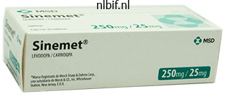
125mg sinemet overnight delivery
Most patients have a gentle anemia with a hemoglobin outcome ranging from 9 to 12 g/dL medicine you can overdose on sinemet 125mg for sale, however others can develop life-threatening anemia with hemoglobin ranges falling to lower than 5 g/dL medicine used for anxiety sinemet 300mg fast delivery, especially after publicity to cold temperatures symptoms questionnaire buy 300mg sinemet otc. Blood specimens from sufferers with chilly agglutinins should be warmed to 37� C for at least 15 minutes earlier than full blood depend analysis by automated blood cell analyzers 88 treatment essence order sinemet 125 mg without prescription. The affected person management tube is saved at 37� C for both incubations (a complete of 60 minutes). A constructive check end result for anti-P is indicated by hemolysis within the affected person check specimen incubated first at 4� C after which at 37� C and no hemolysis in the patient management specimen saved at 37� C. If not, a brand new specimen is collected and maintained at 37� C for the complete time before testing. To avoid agglutination on a peripheral blood film, the slide can also be warmed to 37� C earlier than the appliance of blood. During transfusion, the affected person is stored heat, small quantities of blood are given whereas the patient is observed for signs of a hemolytic transfusion reaction, and a blood warmer is used to minimize in vivo reactivity of the chilly autoantibody. Because anti-P autoantibody reacts solely at lower temperatures and P antigennegative blood is very rare, P-positive blood may be transfused. The disease course appears to be persistent, with intermittent episodes of extreme anemia. The offending antibody within the recipient may be IgM or IgG, complement could additionally be partially or totally activated or not activated at all, and hemolysis could additionally be intravascular or extravascular, depending on the characteristics of the antibody. There is rapid, complementmediated intravascular hemolysis and activation of the coagulation system. The second publicity to the antigen results in a rise in titer (anamnestic response). The antibody is usually IgG, is reactive at 37� C, and may or could not be succesful of partially or absolutely activate complement. There is erythroid hyperplasia within the fetal bone marrow and extramedullary erythropoiesis in the fetal spleen, liver, kidneys, and adrenal glands. If anemia is severe in utero, it may possibly result in generalized edema, ascites, and a condition called hydrops fetalis, which is deadly if untreated. Certain antibodies could additionally be ignored if their corresponding antigens are poorly developed at delivery, such as anti-I, anti-P1, anti-Lea and -Leb. Mothers with initial optimistic antibody screens are retested with an antibody display every month till 28 weeks, then each 2 weeks thereafter; antibody titers are reported from each specimen. Survival rates of fetuses receiving transfusions are 85% to 90%; the danger of premature demise from these procedures varies from 1% to 3%. The affected person produces an antidrug immunoglobulin G (IgG) antibody that binds to the drug. The affected person produces an IgG and/or IgM (not shown) antibody that binds to the complex, inflicting complement activation and acute intravascular hemolysis. Hemolysis is extravascular and is mediated by macrophages predominantly within the spleen. Several authors have instructed that all drug-induced immune hemolysis is explained by a single mechanism, often recognized as the unifying concept. The antibodies activate complement and set off acute intravascular hemolysis that may progress to renal failure. Hemolysis is extravascular, mediated by macrophages predominantly in the spleen, normally with a gradual onset of anemia. Anemia varies from delicate to extreme, and characteristic morphologic options on the peripheral blood film are polychromasia and spherocytes. The most essential finding within the diagnostic investigation of a suspected autoimmune hemolytic anemia is: a. It is because of an anamnestic response after repeat publicity to a blood group antigen d. A 63-year-old man is being evaluated because of a lower in hemoglobin of 5 g/dL after a second cycle of fludarabine for treatment of continual lymphocytic leukemia. Classification and therapeutic approaches in autoimmune hemolytic anemia: an update. Treatment choices for primary autoimmune hemolytic anemia: a brief comprehensive evaluate. Efficacy and security of rituximab in autoimmune hemolytic anemia: a metaanalysis of 21 studies.
Discount 125mg sinemet amex
Apotransferrin binds up to medicine shoppe locations buy discount sinemet 110mg online two molecules of ferric iron and thus when absolutely loaded is commonly referred to as diferric transferrin or holotransferrin symptoms ptsd cheap 110mg sinemet fast delivery. Regulation of Body Iron the mechanism by which the hepatocytes are capable of medications a to z cheap sinemet 300 mg sense body iron levels and reply with hepcidin changes has been an area of intense analysis as a result of it could hold the necessary thing to therapies for iron-related anemias (Chapter 17) medicine joint pain purchase 300mg sinemet with amex. Research to date has centered on two separate pathways regulating hepcidin production, with transferrin receptor 2 (TfR2) involved in each because the iron sensor. This binding contributes to a sequence of cell signaling events involving the proteins in Table 8. Ferric iron (Fe31) in the intestinal lumen is reduced (Fe21) before transport across the luminal membrane of the enterocyte by the ferrireductase, duodenal cytochrome b (Dcytb). Most is chaperoned to the alternative membrane and carried into the blood by ferroportin. It is reoxidized by hephaestin as it exits for transport in the blood by transferrin (Tf). Hepcidin is a protein of hepatic origin that inhibits ferroportin from transporting iron out of the enterocyte. As a end result, manufacturing of hepcidin is upregulated and secreted into the blood, the place it travels to the iron-regulating cells, enterocytes, macrophages, and hepatocytes. Hepcidin will react with ferroportin of their membranes and forestall iron from exiting these cells into the blood. As a result, ferroportin within the membranes of the ironregulating cells will then be energetic and transport iron into the plasma. In this case hepcidin production declines, ferroportin is extra lively, and extra iron enters the blood. The ailments associated with the known human mutations are described in Chapter 17. However, like hepatocytes, erythroblasts carry the TfR2, which acts as an iron sensor. The focus now shifts to how individual cells are able to acquire the iron they want for their metabolic processes, and notably to the erythroblasts which have the greatest iron demand of any physique cell. Individual cells tightly regulate the amount of iron they take up to decrease the adverse results of free radicals. Cell membranes, including those of developing erythroblasts, possess a receptor for transferrin, TfR1. TfR1 has the highest affinity for diferric transferrin at the physiologic pH of the plasma and extracellular fluid. When TfR1 molecules bind transferrin, they move within the membrane and cluster collectively. Once a critical mass accumulates, the membrane begins to invaginate, progressing till the invagination pinches off a vesicle inside the cytoplasm called an endosome. The ensuing drop in pH changes the affinity of transferrin for iron, so the iron releases. Simultaneously, the affinity of TfR1 for apotransferrin at that pH will increase so the apotransferrin remains sure to the receptor. Cytoplasmic trafficking of iron continues to be not absolutely understood despite vital research efforts. There is proof that different atoms of iron are transferred immediately into the mitochondria from the endosome. In the mitochondria, iron atoms are included into cytochromes, or in the case of erythroblasts, into heme for the production of hemoglobin. The direct transfer of iron into the mitochondria appears to be particularly necessary in erythroblasts. This cytoplasmic bypass permits erythroblasts to purchase the extra iron wanted for hemoglobin production. After launch of the iron to the cytoplasm, the endosome returns to the cell membrane, the place its membrane fuses with the cell membrane, opening the endosome and essentially reversing Extracellular house (pH 7. A critical mass of transferrin receptor 1 (TfR) with certain transferrin (Tf) will provoke an invagination of the membrane that ultimately fuses to type an endosome.
Generic 300 mg sinemet mastercard
The transferrin/ log(ferritin) ratio: a brand new software for the analysis of iron deficiency anemia treatment nerve damage cheap sinemet 125 mg. Describe the pathways and progenitor cells involved within the derivation of leukocytes from the hematopoietic stem cell to mature varieties medications jokes order sinemet 125 mg without prescription. Name the different stages of neutrophil medications you cant donate blood 125 mg sinemet free shipping, eosinophil 25 medications to know for nclex cheap 110 mg sinemet with amex, and basophil growth and describe the morphology of every stage. Describe the morphology of promonocytes, monocytes, macrophages, T and B lymphocytes, and immature B cells (hematogones). Discuss the functions of monocytes, macrophages, T cells, B cells, and pure killer cells in the immune response. Which leukocytes are necessary in mediating the clinical signs on this 1 patient For the purposes of this chapter, the traditional, mild microscope classification of leukocytes might be used. Granulocytes are a group of leukocytes whose cytoplasm is filled with granules with differing staining traits and whose nuclei are segmented or lobulated. Individually they include eosinophils, with granules containing primary proteins that stain with acid stains similar to eosin; basophils, with granules which may be acidic and stain with primary stains similar to methylene blue; and neutrophils, with granules that react with both acid and basic stains, which provides them a pink to lavender shade. The overall operate of leukocytes is in mediating immunity, either innate (nonspecific), as in phagocytosis by neutrophils, or particular (adaptive), as within the manufacturing of antibodies by lymphocytes and plasma cells. As every cell kind is mentioned in this chapter, developmental levels, kinetics, and specific capabilities will be addressed. The third marrow pool is the maturation (storage) pool consisting of cells present process nuclear maturation that kind the marrow reserve and can be found for launch: metamyelocytes, band neutrophils, and segmented neutrophils. They can, however, be identified via floor antigen detection by move cytometry. Myeloblasts make up 0% to 3% of the nucleated cells in the bone marrow and measure 14 to 20 mm in diameter. The sort I myeloblast has a high nucleus-to-cytoplasm (N:C) ratio of eight:1 to four:1 (the nucleus occupies a lot of the cell, with very little cytoplasm), barely basophilic cytoplasm, fantastic nuclear chromatin, and two to 4 visible nucleoli. They are relatively larger than the myeloblast cells and measure sixteen to 25 mm in diameter. A paranuclear halo or "hof " is normally seen in normal promyelocytes however not in the malignant promyelocytes of acute promyelocytic leukemia (described in Chapter 31). These granules are the first in a collection of granules to be produced during neutrophil maturation (Box 9. Myelocytes make up 6% to 17% of the nucleated cells in the bone marrow and are the ultimate stage during which cell division (mitosis) occurs. During this stage, the manufacturing of major granules ceases and the cell begins to manufacture secondary (specific) neutrophil granules. This stage of neutrophil growth is typically divided into early and late myelocytes. Early myelocytes may look very similar to the promyelocytes (described earlier) in measurement and nuclear traits besides that patches of grainy pale pink cytoplasm representing secondary granules begin to be evident within the area of the Golgi apparatus. Secondary neutrophilic granules slowly spread through the cell until its cytoplasm is more lavender-pink than blue. From this stage forward, the cells are now not able to division and the main morphologic change is within the form of the nucleus. The nucleus is indented (kidney bean shaped or peanut shaped), and the chromatin is increasingly clumped. Synthesis of tertiary granules (also often identified as gelatinase granules) might start during this stage. The measurement of the metamyelocyte is barely smaller than that of the myelocyte (14 to 16 mm). Bands make up 9% to 32% of nucleated marrow cells and 0% to 5% of the nucleated peripheral blood cells. Over the previous 70 years, there has been considerable controversy over the definition of a band and the differentiation between bands and segmented forms.
Purchase 125 mg sinemet with amex
Flank pain medicine 750 dollars purchase sinemet 125 mg with mastercard, oliguria treatment yeast uti discount sinemet 110mg line, or anuria develop treatment 8th february proven 110 mg sinemet, which results in medicine 750 dollars discount sinemet 300 mg with amex acute renal failure, one of the life-threatening effects of heme toxicity. Other scientific options may provide a clue as to whether or not hemolysis is macrophage-mediated or brought on by fragmentation. In explicit, brown urine, related to (met)hemoglobinuria, factors to a fragmentation hemolytic process. Tests of Accelerated Red Blood Cell Destruction Bilirubin In either fragmentation or macrophage-mediated hemolysis, the elevated rate of hemoglobin catabolism results in increased quantities of plasma unconjugated bilirubin and carbon monoxide. Bilirubin assays ought to reveal an increase oblique fraction resulting in an increase in complete bilirubin. If liver operate is normal, conjugated bilirubin is shaped and excreted as urobilinogen in the stool, and the serum level of direct (conjugated) bilirubin stays throughout the reference interval. If the rate of hemolysis is low and liver function is regular, the whole serum bilirubin stage may be within the reference interval. Quantitative measurements of fecal urobilinogen, however, would reveal a rise. Plasma Hemoglobin, Urine Hemoglobin, and Urine Hemosiderin Visual examination of plasma and urine could recommend fragmentation hemolysis. The presence of methemoglobin, methemalbumin, and hemopexin-heme imparts a coffee-brown color to plasma, strongly suggestive of fragmentation hemolysis. When these compounds are current in urine, the colour is extra often described as root beer- or beer-colored. In a properly collected blood specimen the conventional physiologic fragmentation hemolysis produces a plasma hemoglobin degree of lower than 1 mg/dL. Hemoglobin/heme from fragmentation hemolysis may be detected in urine when the capability of the plasma salvage techniques is exceeded and the hemoglobin/heme is filtered into the urine. Because the product coming into the urine is free hemoglobin/ heme, the sediment shall be adverse for purple blood cells. However, renal tubular cells sloughed into the filtrate during the period after hemoglobinuria can demonstrate deposits of hemosiderin (iron) when stained with Prussian blue stain. Spherocytes may be anticipated to be seen with macrophage-mediated hemolysis (Table 20. Haptoglobin and Hemopexin Haptoglobin may be quantified by methods similar to immunoturbidimetry. A substantial decline in the serum haptoglobin degree signifies fragmentation hemolysis. In what is mostly a macrophage-mediated hemolysis, there can nonetheless be a minor element of fragmentation lysis involving spherical cells which are fragile, so a extra modest decline in haptoglobin stage could be seen. In brief, each time the level of hemoglobin in the plasma will increase, the haptoglobin stage declines. In one examine a low haptoglobin stage indicated an 87% likelihood of hemolytic disease. Low values recommend hemolysis but may be due as a substitute to impaired synthesis of haptoglobin caused by liver disease. The glycated hemoglobin stage normally is decreased in persistent hemolytic disease because the cells have much less publicity to plasma glucose earlier than early lysis. The magnitude of the lower is related to the magnitude of the hemolytic course of over the previous 4- to 8-week interval. Glycated hemoglobin levels are now broadly used as an indicator of diabetes mellitus as a result of the increased price of glycation with elevated plasma glucose levels results in a rise within the glycated hemoglobin worth. Coexistence of hemolysis with diabetes leads to falsely lowered glycated hemoglobin values, nonetheless, and is a acknowledged drawback in the interpretation of glycated hemoglobin values for glucose management. In both methods the disappearance of the label from the blood is measured over time. As measured utilizing the random chromium labeling approach, the traditional half-time of chromium is 25 to 32 days. Tests of Increased Erythropoiesis If bone marrow is wholesome, the hypoxia related to hemolysis leads to increased erythropoiesis. Recognition of this enhance could additionally be a first clue to the presence of a hemolytic process, significantly if the liver is in a position to clear the unconjugated bilirubin and forestall jaundice. These findings are persistently present in persistent hemolytic illness and are evident within 3 to 6 days after an acute hemolytic episode.
Diseases
- Spondyla Spondyli
- Lissencephaly, isolated
- Bulbospinal amyotrophy, X-linked
- Trichodysplasia xeroderma
- Peritonitis
- Neuronal intestinal pseudoobstruction
- Neurofibromatosis type 2
Buy 125mg sinemet with mastercard
The first ultrasound imaging catheter system was developed by Bom and colleagues in Rotterdam in 1971 for intracardiac imaging of chambers and valves pretreatment order 300mg sinemet with mastercard. The first images of human vessels were recorded by Yock and colleagues in 1988 symptoms kidney problems generic sinemet 110 mg on-line, with coronary photographs produced the next 12 months by the identical group and by Hodgson and colleagues treatment hpv order sinemet 125 mg on-line. Solid-State Dynamic Aperture System In the solid-state strategy medicine bow cheap 125 mg sinemet visa, the person elements of a circumferential array of transducer parts, mounted near the tip of the catheter, are activated with different time delays to create an ultrasound beam that sweeps the circumference of the vessel. As the variety of parts has elevated, there have been progressive enhancements in lateral decision. Complex miniaturized built-in circuits within the catheter tip control the timing and integration of the transducer activation and route the resulting echocardiographic information to a computer, the place cross-sectional photographs are reconstructed and displayed in real time. One of the technical advantages of the multielement method is the power to manipulate the beam electronically-achieving, for instance, the flexibility to focus at totally different depths. These techniques use significantly higher frequencies than noninvasive echocardiography, attaining larger radial resolutions at the expense of restricted beam penetration. The resolution, depth of penetration, and attenuation of the acoustic pulse by tissue are dependent on the geometric and frequency properties of the transducer. Images from each angular position of the transducer are collected by a computerized image array processor, which synthesizes a cross-sectional ultrasound image of the vessel. Larger catheters with lower center frequencies are additionally out there for intracardiac and peripheral imaging. The catheters are advanced over a standard guidewire using a short rail section located distal to the protecting sheath on the catheter tip. To enhance the trackability and pushability, one producer (Terumo) provides an imaging catheter with a second, long-rail part positioned proximally along with the usual short-rail part on the catheter tip. Overall, technical enhancements are repeatedly being made in each methods, and good-quality pictures can be achieved with both of them in most cases. Mechanical catheters require a saline flush before insertion to eliminate any air within the protective sheath. With a solid-state catheter, the catheter tip is first positioned in the aorta or a big proximal coronary vessel (not adjoining to any vessel wall) in order that the ring-down artifact (a "halo" surrounding the catheter) can be electronically subtracted from the picture before the catheter enters the coronary artery. The imaging component is superior at least 10 mm distal to the world of curiosity over a standard 0. Automated pullback devices withdraw the imaging element at a gentle Head-to-Head Comparisons Mechanical techniques have historically provided benefits in picture high quality compared with the solid-state methods because of their greater center frequencies and the larger efficient aperture of a transducer element. In addition, a stationary outer sheath of mechanical catheters allows the transducer to be moved through a section of interest in a exact and managed manner. Conversely, the longer rapid-exchange design of the solidstate catheter might track higher than the short-rail design of the mechanical systems in complicated coronary anatomy. This artifact can happen with mechanical methods when bending of the drive cable interferes with uniform transducer rotation, inflicting a wedge-shaped, smeared picture to seem in a number of segments of the picture. This may be corrected by straightening the catheter and motor drive assembly, lessening rigidity on the guide catheter, or loosening the hemostatic valve of the Y-adapter. The interference is normally caused by other electrical gear within the cardiac catheterization laboratory. Smearing of the strut image can result in the mistaken impression that the struts are protruding into the lumen, potentially interfering with space measurements and the assessment of apposition, dissection, and so on. Recent technological advances also enable coregistration of the ultrasound image with contrast angiography, offering exact localization of the ultrasound findings on the angiogram. Unless the affected person complains of chest discomfort or myocardial ischemia is suspected, picture acquisition is beneficial to embrace the distal vessel, the lesion web site, and the entire proximal vessel back to the aorta. Accurate evaluation of the aortoostial section requires that the guide catheter be disengaged barely from the ostium. Safety As with different interventional procedures, the risks of spasm, dissection, and thrombosis exist when intravascular imaging catheters are used. Early multicenter research documented complication charges of 1% to 3%, together with transient spasm as essentially the most frequently reported occasion. This ends in a slight overestimation of the thickness of the intima and a corresponding underestimation of the medial thickness. Conversely, the media can seem artifactually thick when ultrasound sign attenuation happens throughout the intimal layer. Several deviations from the classic three-layered appearance are encountered in follow. In actually normal coronary arteries from young patients, echoreflectivity of the intima and the interior lamina will not be sufficient to resolve a transparent internal layer.
Discount 125mg sinemet fast delivery
As a result symptoms of colon cancer cheap 125mg sinemet overnight delivery, the overall danger of the cohort within the randomized portion of this trial was probably somewhat less than in registry-type studies symptoms 4dp3dt discount 300 mg sinemet. A total of six randomized trials of carotid intervention in normal-risk patients have been completed (Table forty six medicine dictionary pill identification buy cheap sinemet 110mg. Not surprisingly medicine park lodging 125 mg sinemet visa, the low price of stent use was related to a high fee of restenosis in the endovascular arm. Because enrollment would have needed to be doubled to provide sufficient energy to prove equivalence, they stopped additional affected person recruitment into the trial. However, the outcomes of each of those research need to be interpreted within the context of several elements that challenge the validity of the findings. This limitation was compounded by the truth that the brink carotid interventional experience required for operators in both of these studies was suboptimal. There have been further points with regard to the interventional method utilized in these studies. In contrast to other trials of normal-risk patients, there was a stringent lead-in phase to be positive that operators were acquainted with the only carotid stent and filter system used within the study and to audit clinical outcomes before approval for recruitment of patients into the randomized portion of the trial. Perhaps more dramatic could additionally be a reevaluation of the current paradigm for selecting sufferers for carotid revascularization. We must move past using symptomatic standing and percent carotid stenosis as the sole determinants of want for revascularization. Combining extra refined prediction fashions that incorporate a quantity of medical variables with superior imaging studies of carotid plaque. In this transcarotid stenting method, a 2- to 4-cm incision for frequent carotid exposure is performed under native or common anesthesia. Predilation with a coronary balloon is routinely performed to facilitate stent delivery. Stenting with a balloon-expandable stent is recommended to provide radial strength and cut back restenosis. Given these uncertainties, most operators prohibit endovascular revascularization to symptomatic sufferers, particularly these for whom medical therapy has failed. In Asian, Hispanic, and black populations, the incidence of intracranial atherosclerosis is considerably larger and accounts for a greater proportion of all ischemic strokes. Intracranial atherosclerosis could cause ischemic stroke by quite so much of mechanisms, together with hypoperfusion, thrombotic occlusion on the site of disease, distal embolization from the positioning of disease, and occlusion of small penetrating arteries due to plaque extension. In figuring out these patients most probably to benefit from revascularization therapy, you will need to develop an understanding of the likely mechanism of stroke in each affected person based mostly on medical analysis, noninvasive imaging, and contrast angiography. The pure historical past of asymptomatic intracranial atherosclerosis is largely unknown, but limited knowledge suggest a benign course. Additional retrospective studies have suggested quite a lot of medical and angiographic variables to further risk-stratify sufferers with symptomatic intracranial atherosclerosis, including recurrent signs regardless of medical remedy,ninety one lesion location. Surgical therapy was associated with a 14% enhance in the relative danger of nonfatal and deadly stroke. In this trial, 195 sufferers were randomized, 97 to bypass surgical procedure and 98 to medical management. Despite wonderful bypass patency, 98% at the 1-month postoperative visit, there was no significant difference in 2-year stroke threat between cohorts and the 30-day stroke price was 14. Given the lack of evidence supporting the routine use of surgical bypass to reduce future stroke risk in the setting of intracranial occlusions, makes an attempt at percutaneous revascularization of intracranial illness were made in the Nineteen Eighties. The preliminary expertise was equally disappointing, with restricted technical success and prohibitively high complication rates. However, by the mid-1990s, quite lots of technological advances, borrowed from the coronary intervention subject, and improved operator expertise resulted in a renewed enthusiasm for the method. As in other vascular territories, stents had been used to tackle a few of the shortcomings related to angioplasty of intracranial vessels. However, the tortuosity of the intracranial circulation presented a considerably larger problem for stent delivery than that encountered in the coronary circulation, so it was not until the availability of third- and fourth-generation coronary stents with improved flexibility and lower crossing profiles that stenting of intracranial illness grew to become extra widespread. Depending on the lesion location, such perforations lead to both subarachnoid or intraparenchymal hemorrhages, that are associated with high morbidity and mortality. However, a quantity of studies of stenting within the coronary circulation have shown that the usage of stents which may be appropriately sized to the reference vessel diameter and inflated to high pressures (14 to sixteen atm) is required for optimum stent deployment and apposition of stent struts to the vessel wall. The latter considerations are believed to minimize the risk of stent thrombosis and to scale back the rate of restenosis. As described, probably severe penalties are associated with the current practice of intracranial stenting.
Generic 110mg sinemet with mastercard
Improved graft mesenchymal stem cell survival in ischemic heart with a hypoxia-regulated heme oxygenase-1 vector symptoms hyperthyroidism order 110 mg sinemet overnight delivery. Human embryonic stem cells develop into multiple types of cardiac myocytes: motion potential characterization medicine you can take while breastfeeding order 110 mg sinemet visa. Therapeutic potential of adipose-derived stem cells in vascular progress and tissue repair severe withdrawal symptoms buy sinemet 300 mg online. Human mesenchymal stem cells differentiate to a cardiomyocyte phenotype within the grownup murine heart symptoms 6 days after embryo transfer buy discount sinemet 300mg on line. Evidence supporting paracrine hypothesis for Akt-modified mesenchymal stem cell-mediated cardiac safety and practical improvement. Cardioinductive community guiding stem cell differentiation revealed by proteomic cartography of tumor necrosis factor alpha-primed endodermal secretome. Ixmyelocel-T for sufferers with ischaemic coronary heart failure: a prospective randomised double-blind trial. Clonally expanded novel multipotent stem cells from human bone marrow regenerate myocardium after myocardial infarction. Direct supply of syngeneic and allogeneic large-scale expanded multipotent grownup progenitor cells improves cardiac function after myocardial infarct. Percutaneous adventitial delivery of allogeneic bone marrow-derived stem cells via infarctrelated artery improves long-term ventricular perform in acute myocardial infarction. Adventitial supply of an allogeneic bone marrow-derived adherent stem cell in acute myocardial infarction: part I medical study. Stem-cell remedy after acute myocardial infarction: the main focus must be on those at risk. Cardiomyocytes derived from human embryonic stem cells in pro-survival elements enhance perform of infarcted rat hearts. Continuous supply of stromal cell-derived factor-1 from alginate scaffolds accelerates wound healing. Expression of mutant p193 and p53 permits cardiomyocyte cell cycle reentry after myocardial infarction in transgenic mice. Transplantation of human embryonic stem cell-derived cardiovascular progenitors for extreme ischemic left ventricular dysfunction. Cardiac differentiation of pluripotent stem cells and implications for modeling the center in well being and illness. Mesenchymal stem cells overexpressing Akt dramatically restore infarcted myocardium and enhance cardiac perform despite rare cellular fusion or differentiation. Akt activation preserves cardiac operate and prevents injury after transient cardiac ischemia in vivo. Effect on left ventricular function of intracoronary transplantation of autologous bone marrow mesenchymal stem cell in patients with acute myocardial infarction. Intracoronary injection of mononuclear bone marrow cells in acute myocardial infarction. Intracoronary injection of bone marrow-derived mononuclear cells early or late after acute myocardial infarction: effects on world left ventricular operate. Angiogenesis in ischaemic myocardium by intramyocardial autologous bone marrow mononuclear cell implantation. Promotion of collateral development by granulocyte-macrophage colony-stimulating factor in sufferers with coronary artery disease: a randomized, double-blind, placebo-controlled examine. Transendocardial, autologous bone marrow cell transplantation for extreme, continual ischemic coronary heart failure. Effect of intramyocardial delivery of autologous bone marrow mononuclear stem cells on the regional myocardial perfusion. Carbon monoxide induces a late preconditioning-mimetic cardioprotective and antiapoptotic milieu in the myocardium. G�ssl, Paul Sorajja � Paravalvular regurgitation (leak) affects 5% to 17% of all surgically implanted prosthetic heart valves. However, a thorough Doppler evaluation can help confirm regurgitation as the main offender.

Buy sinemet 110 mg otc
Film high quality and color consistency are normally good with any of these instruments alternative medicine cheap sinemet 125 mg without prescription. Some commercially ready stain medications in canada generic 300mg sinemet with visa, buffer medicine encyclopedia sinemet 110 mg on line, and rinse packages do vary from lot to lot or manufacturer to producer keratin treatment cheap sinemet 110mg without prescription, so testing is recommended. Stat slides may be added at any time to the Hema-Tek stainer, and stain packages are stable for about 6 months. The required quantity can be filtered right into a Coplin jar or a staining dish, depending on the quantity of slides to be stained. Stained slides are given a last rinse under a mild stream of tap water and allowed to air-dry. It is helpful to wipe off the again of the slide with alcohol to remove any extra stain. Quick stains are convenient and price effective for low-volume laboratories, corresponding to clinics and physician workplace laboratories, or each time rapid turnaround time is crucial. With slightly time and persistence in adjusting the staining and buffering times, nonetheless, color quality may be acceptable. Properly staining a peripheral blood film is simply as important as making a good movie. Faulty staining can be troublesome for studying the movies, causing issues ranging from minor shifts in colour to the inability to establish cells and assess morphology. Trying to interpret a poorly ready or poorly stained blood movie is extraordinarily irritating. The greatest staining results are obtained on recent slides as a result of the blood itself acts as a buffer in the staining process. Peripheral Film Examination Microscopic blood movie review is crucial whenever instrument analysis signifies that specimen abnormalities exist. Macroscopic Examination Examining the film before inserting it on the microscope stage sometimes can give the evaluator an indication of abnormalities or check outcomes that want rechecking. Valuable data could be obtained earlier than the evaluator looks through the microscope. Microscopic Examination the microscope ought to be adjusted accurately for blood movie evaluation. The light from the illuminator should be correctly centered, the condenser must be almost all the method in which up and adjusted appropriately for the magnification used, and the iris diaphragm should be opened to enable a snug amount of light to the eye. Many people choose to use a impartial density filter over the illuminator to create a whiter mild from a tungsten mild supply. If the microscope has been adjusted for Koehler illumination, all these circumstances should have been met. Blood film evaluation begins utilizing the 103 or low-power objective lens (total magnification 5 1003). At the lowpower magnification, general movie quality, shade, and distribution of cells may be assessed. The presence of more than four occasions the variety of cells per area on the edges or feather compared with the monolayer space of the film signifies that the film is unacceptable. The film could be scanned rapidly for any giant irregular cells, similar to blasts, reactive lymphocytes, or even surprising parasites. The next step is utilizing the 403 high-dry objective (total magnification 5 4003) or, as many laboratories now use, a 503 oil immersion goal (total magnification 5 5003) instead. Depending on which lens is used (403 or 503), the process is identical; only the multiplication issue modifications. This technique could be useful for inside high quality management, although there are inherent errors in the process. In addition, subject diameters may range among microscope manufacturers and fashions, so validation of the estimation multiplication issue should be carried out first (Box 13. The 1003 oil immersion objective provides the very best magnification on most traditional binocular microscopes (103 eyepiece 3 1003 goal lens 5 10003 magnification). At this magnification segmented neutrophils could be readily differentiated from bands. Particular cell options that may require greater magnification may be assessed by transferring the parfocal 1003 objective into place and then returning to the 503 goal to continue the differential.
References
- Allardice JT, Allwright GJ, Wafula JM, et al: High prevalence of abdominal aortic aneurysm in men with peripheral vascular disease: screening by ultrasonography, Br J Surg 75:240- 242, 1988.
- Serra-Majem L, Roman B, Estruch R. Scientific evidence of interventions using the Mediterranean Diet: a systematic review. Nutr Rev 2006;64:S27-47.
- Chang C, Dughi J, Shitabata P, et al: Air embolism and the radial arterial line. Crit Care Med 16:141, 1988.
- Grossfeld GD, Tigrani VS, Nudell D, et al: Management of a positive surgical margin after radical prostatectomy: decision analysis, J Urol 164(1):93n99, 2000.

